References


Borrelia Burgdorferi: Symptoms and Treatment. (n.d.). Borrelia Burgdorferi. Borrelia Burgdorferi: Symptoms and Treatment.
Retrieved from http://borreliaburgdorferi.org/.

Ghiasvand, A. (2011, Nov. 11). Life Cycles & Reproduction. [Awesome Inc.]. 
Retrieved from http://borrelia-burgdorferi.blogspot.ca/2011/11/life-cycles-reproduction.html.

Hunt, Richard. (2010, April 19). Parasitology Chapter Seven Part Two Ticks. Microbiology and Immunology On-line University of South Carolina School of Medicine. 
Retrieved from http://pathmicro.med.sc.edu/parasitology/ticks.htm.

American Society for Microbiology. (2013, March 25). Motility is crucial for the infectious life cycle of Borrelia burgdorferi. American Society for Microbiology: Infection and Immunity. Retrieved from http://iai.asm.org/content/early/2013/03/19/IAI.01228-12.abstract.

Kung F., Anguita J., & Pai U. (n.d.). Borrelia Burgdorferi and Tick Proteins Supporting Pathogen Persistence in the Vector. Medscape News. Retrieved from http://www.medscape.com/viewarticle/776862.


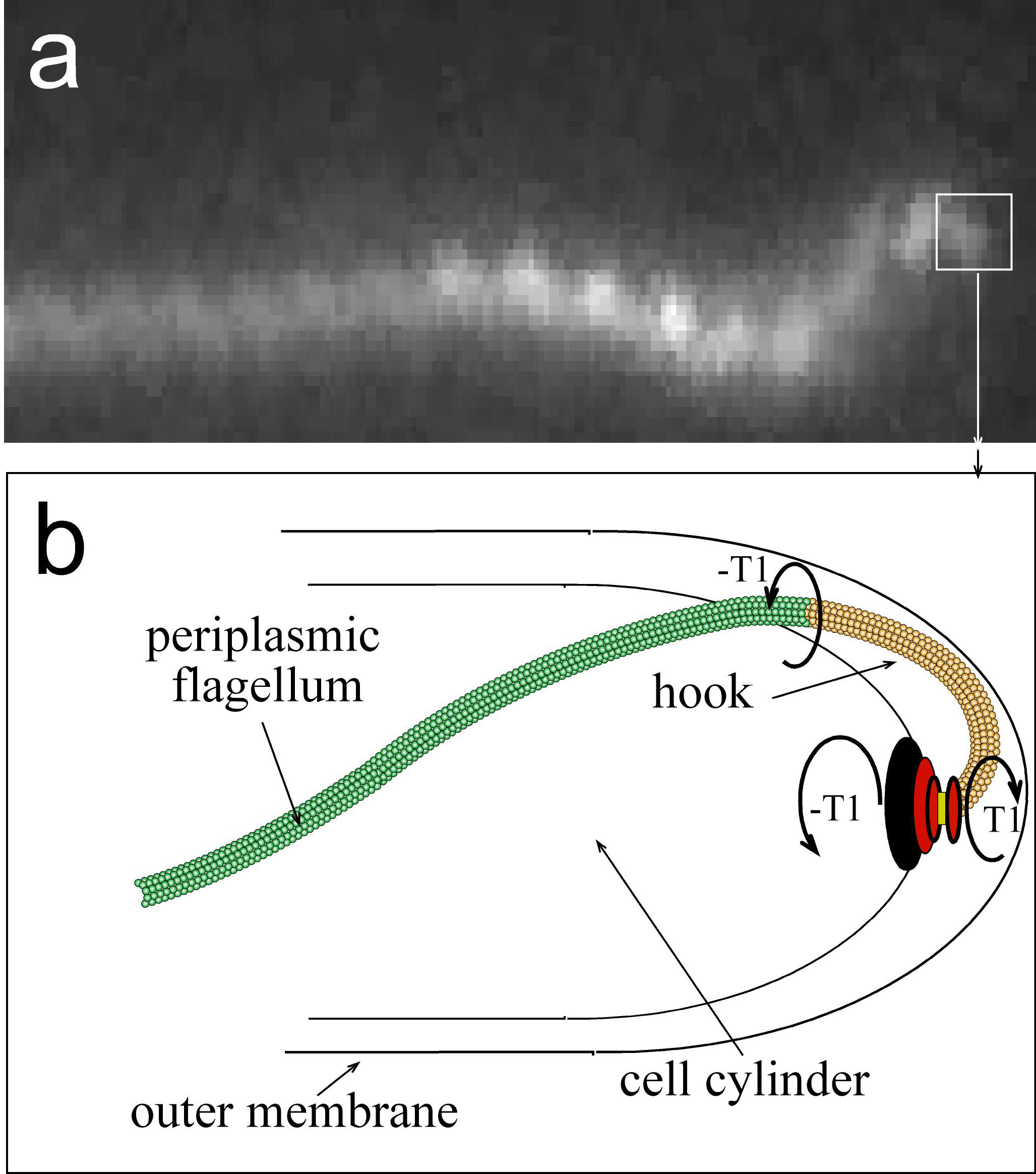
Sal M. S., Li C., Motalab. M. A., Shibata S., Aizawa S., & Charon N. W. (2008, Jan. 11). Borrelia burgdorferi Uniquely Regulates Its Mobility Genes and Has an Intricate Flagellar Hook-Basal Body Structure. National Center for Biotechnology Information, U.S. National Library of Medicine. Retried from http://www.ncbi.nlm.nih.gov/pmc/articles/PMC2258876/.

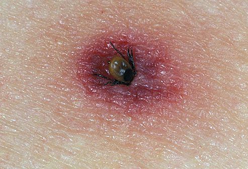
Interactive Medical Media LLC. (n.d.). Tick Bites. [Photograph]. Retrieved from http://www.webmd.com/skin-problems-and-treatments/picture-of-tick-bites.

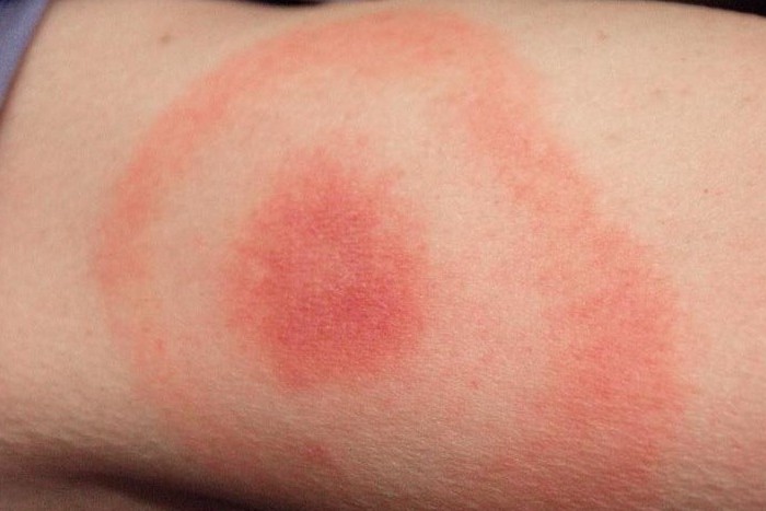
Gathany J. (2010, Dec. 8). Lyme Disease Rash Picture. [Photograph]. Retrieved from http://hardinmd.lib.uiowa.edu/cdc/lymedisease6.html.

VanDyk J. (1996, June 12). Ixodes Scapularis, the Black-Legged or Deer Tick. [Photograph]. Retrieved from http://www.ent.iastate.edu/imagegallery/ticks/iscapall4wd.html.

Stanek, G. (2012). Lyme borreliosis. [Microscopic Diagram]. 
Retrieved from http://www.sciencedirect.com/science/article/pii/S0140673611601037.

Bacmap Genome Atlas. (n.d.). Borrelia burgdorferi B31. [Microscopic Photograph]. Retrieved from http://bacmap.wishartlab.com/organisms/19.

Rosa, P. A., Tilly K., & Stewar, P. E. (2005). Lyme Disease. [Diagram].
Retrieved from http://www.nature.com/nrmicro/journal/v3/n2/box/nrmicro1086_BX1.html.

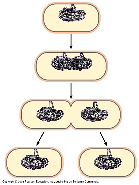
Biology Exams 4 U. (2013). Difference between prokaryotic and eukaryotic chromosome. [Diagram].
Retreived from http://www.biologyexams4u.com/2012/11/difference-between-prokaryotic-and.html.

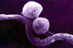
Micro*scope. (2006). Mixed Population. [Microscopic Photograph].
Retrieved from http://starcentral.mbl.edu/microscope/portal.php?pagetitle=assetfactsheet&imageid=755. 

Vojdani A., Hebroni F., Raphael Y., Erde J., & Raxien B. (2007). Novel Diagnosis of Lyme Disease: Potential for CAM Intervention. [Diagram]. 
Retrieved from http://openi.nlm.nih.gov/detailedresult.php?img=2722197_nem138f1&req=4.

Kumaran D., Eswaramoorthy S., Luft B. J., Koide S., Dunn J. J., Lawson C. L., & Swaminathan S. (2001). Crystal structure of outer surface protein C (OspC) from the Lyme disease spirochete, Borrelia burgdorferi. [Diagram].
Retrieved from http://www.nature.com/emboj/journal/v20/n5/fig_tab/7593602a_F2.html.

AMI Health. (2010). Lyme Disease Treatment. [Poster]. 
Retrieved from 
http://www.amihealth.com/lyme-disease-treatment.html.

(n.d.) Spirochete Morphology and Motility. [Photograph & Diagram] Retrieved from http://www.physics.arizona.edu/~wolg/research.html.

American Lyme Disease Foundation. (2010). Deer Tick Ecology. [Diagram]. Retrieved from
http://www.aldf.com/deerTickEcology.shtml.

Relationship with Humans


B. burgdorferi has a highly relevant connection with humans. It is well-known for causing the most common vector-borne bacterial disease in the world, 
with over 20 000 infections per year: the Lyme disease.


Fig.22. A Sign Warning of
the Presence of Ticks

Lyme disease is the most common in areas where the transmitter of the disease, the Ixodes ticks are found in: 
wooded ares in the northern hemisphere.


Humans are secondary hosts of B. burgdorferi.
The only way humans can be infected by B. burgdorferi is when a carrier of this pathogen accidentally attach to and bite humans, while searching for other mammals, such as a deer.

Fortunately, transmission from human to human is unknown, not even during pregnancy or through breast milk: Lyme disease is not contagious.


Fig.23. A Tick Attached
to Human Skin
In order for B. burgdorferi to be transmitted, the ticks need to be attached to the host skin for 24 to 48 hours, with the success of the transmission increasing with the tick attachment duration.


Lyme disease can be divided into three distinct stages.


Symptoms:

Fig.24. An Erythema Migran
Stage I: Erythema chronicum migrans (a circular red rash at the site of the tick bite, resembling a bull’s eye) and flu-like symptoms: fatigue, fever,headache, muscle and joint aches, and malaise,  a feeling of “discomfort or uneasiness”.

If the symptoms are not recognized and the infection is left untreated, the disease moves on to its second stage.

Stage II: facial palsy (or paralysis), meningitis (inflammation of the protective membranes of the brain and spinal cord), extreme joint and muscle pain, and palpitation (abnormality in heartbeat)

After several months of disregard, the Stage III beings.

Stage III: arthritis (a type of joint disorder) accompanied by severe joint pain and swelling, vision problems


Treatment:
A vaccine for human use is currently unavailable. 

Fig.25, Treatment for Lyme Disease
Involves Taking Antibiotics.
However, in the early stage of this disease, a few-weeks-long treatment involving antibiotics like amoxicillin or doxycycline is available.

In the late stages of this disease, treatment usually takes months of various antibiotics and other drugs. Being a Gram-negative bacterium with an impenetrable wall, it is highly resistant to antibodies and antibiotics.

Therefore it is crucial for early diagnosis and subsequent treatment.


Diagnosis:
Tick bites are usually painless, and symptoms of Lyme disease during its early stages of Lyme are easily mistaken for typical flus or rashes; therefore, it isn't uncommon for Lyme disease to be left untreated for months before people become suspicious of an infection. 

Diagnosis of this infection is done through a blood test. 
The presence of antibodies will indicate that the body has been infected by B. burgdorferi. Antibodies are proteins created by the body’s immune system to fight foreign substances.

Lyme Disease Treatment
Fig.26. Watch Out for
InfectiousTicks!

Prevention:
The best way to prevent Lyme disease is to protect the body from Ixodid ticks, by minimizing skin exposure when in wooded or grassy areas, which is where the ticks are the most commonly found.


Currently, scientists are trying to figure out the pathogenesis of B. burgdorferi, the mechanism that causes the Lyme disease
However, due to its specific nutritional requirements, 
the bacterium is difficult to culture in vitro. 

Reproduction


The species, B. burgdorferi, has never been found naturally outside of a host. This indicates that it requires a host body in order to reproduce itself.
Fig.20. Stages in
Binary Fission


Upon entering the host, B. burgdorferi reproduces asexually mainly through binary fission.

However, it also uses other methods of reproduction, one of which involves the formation of a cyst. 
A cyst is a sac containing air and some fluids enclosed by its own membrane that forms when a single B. burgdorferi cell curls into a cocoon around itself. A cyst, from which young B. burgdorferi forms is created and released.

Fig.21. The Formation of
a B. burgdorferi's Cyst

Another mechanism of reproduction that this bacterium utilizes is the formation of buds, which then can eventually turn into cysts. 

The bursting of the B. burgdorferi cell that contain these underdeveloped forms is how they are released.


B. burgdorferi has a unique ability to reproduce wherever and whenever as needed, no matter the surroundings, while also simultaneously protecting itself from its hosts’ immune system.


Its ability to reproduce is one of the many contributions to its success in invading host organisms.

Life Cycle

Fig.16. Ixodid Tick and
B. burgdorferi

B. burgdorferi circulates between lxodes ricinus ticks and a large variety of mammalian hosts, usually small rodents, in a zoonotic cycle 
(zoonosis: an infectious disease that can be transmitted between species).


The lifecycle of B. burgdorferi is closely related to that of its primary host, the Ixodes ticks.
The ticks are the bacterium’s vector: an organism that has been previously infected by B. burgdorferi and acts as its carrier and transmitter. 
The infected mammals are the reservoir, a “holding area” for these spirochetes.


Fig.17. Seasonal 2-Year Lifecycle
of a Deer Tick
The hatching of a tick larva in early summer marks the start of this pathogenic species’ lifecycle. 
The larva attaches to and bites into a host that has been previously infected by B. burgdorferi, allowing the bacterium to enter the tick’s bloodstreams.
B. burgdorferi continues to flow through the blood to all parts of the tick’s body, such as the heart, brain, muscles, bones, and eventually into its gut.

Once in the tick’s gut, B. burgdorferi attaches to the lining of the gut using its surface proteins, creating colonies of the bacterium. 
It then penetrates the gut wall and multiplies in the spaces between the tick’s cells. These colonies of B. burgdorferi remain in this state until the next time the tick feeds.

 When it does, the B. burgdorferi reactivates and enters the tick’s hemolymph to its salivary glands. 
It is through the tick’s saliva that the spirochetes can be transmitted to a new host.
Once in a mammalian host, the bacterium remains in it for the rest of its life.


Fig.18. Relative Sizes of Ixodes Ticks
at Different Stages
An Ixodes tick’s two-year long life cycle can be divided into three stages: larva, nymph, and adult. At each stage, the tick must take a “meal” and it is during this time that B. burgdorferi is transmitted between species.


After growing into a nymph from a larva, the tick will detach itself from the host. The nymph stage is the dormant stage of the tick’s life, and it is in this nymph stage that it remains through the winter. 
The next spring, the adult tick reproduces and 
once again a B. burgdorferi lifecycle begins.


Fig.19. The Lifecycle of a B. burgdorferi and its Interconnected Organisms


Although B. burgdorferi is known to negatively affect some animals, including humans, 
through evolution, their primary hosts, the Ixodid ticks have developed tolerance to this pathogen. 
The ticks’ ability to withstand B. burgdorferi allows the two organisms to form a stable symbiotic relationship.

Movement


B. burgdorferi’s periplasmic flagella act as a motor.

Fig.14. Mechanism Behind
B. burgdorferi's Motion
With a “hook” that spins on its axis, the bacterium’s flagella rotate, creating a backward, “corkscrew” motion. This rotation of the flagella enables B. burgdorferi to propel itself and swim in both low and highly viscous fluids and materials.

The flagella are located in the periplasmic space: between the cytoplasmic membrane and the outer membrane. 
These two membranes allow the flagella to “drive the rest of the cell around the long axis” and to create a motion capable of penetrating complex tissues within and between different hosts.

They also offer the bacterium protection against its host’s immune system. 
The flagella allow it to outrun the host’s immune cells.


Mortility of B. burgdorferi is a very significant part of its success in invading other organisms.

 Fig.15. B. burgdorferi in Motion
From the tick gut, B. burgdorferi invades the tick’s dermis, which is a layer of skin between the epidermis (the outermost) layer and the hypodermis (lowermost) layer.
 From the dermis, B. burgdorferi disperses throughout the tick’s body to its internal organs and tissues, including myocardium (muscular tissues in the heart), Synovial fluid (found in humans’ joints), heart, and the neurological system.

Nutrition


The ecological niche of this bacterium is heavily dependent on the Ixodid ticks. 
Not only are they the primary hosts of B. burgdorferi, 
they act as a carrier of the pathogen, allowing the bacterium to attach to a new mammalian host.

Borrelia Burgdorferi
Fig.12. B. burgdorferi Attached to
Host Organisms
B. burgdorferi’s main source of nutrition is parasitic nutrition. 
Parasitic nutrition is a type of heterotrophic nutrition where a parasite that lives on the surface or inside of another host organism feeds off of the nutrients from the digested food.

This endoparasitic bacterium lacks genes required for cellular synthesis and metabolism, 
which is why it needs a host to survive.


B. burgdorferi's capability of changing its nourishment sources from the mammalian blood, such as glucose and other carbohydrates, to the ticks’ hemolymph 
(a fluid in the circulatory system of arthropods) allow the species to survive in the bodies of different hosts.

Fig.13. B. burgdorferi's Enzyme
has been Replaced By Manganese
 
B. burgdorferi is one of the few bacteria that don’t require iron in its diet. (In fact, it was the first organism to live without iron.) 
Its iron-sulfur cluster enzymes are replaced by those of manganese.

Habitat


B. burgdorferi is an endoparasite
An endoparasite is a parasite that lives inside its host. Therefore, its habitat will be areas in which its hosts are abundant.


Figure.10. An Example of a Habitat
in which Ixodid Ticks can be Abundant.
The Ixodes ticks are the most dominant organisms in which B. burgdorferi live; 
and, they’re most commonly found in wooded areas in the northern hemisphere, such as North America, and parts of Europe and Asia.


Fig.11. Global Distribution of Ixodes Ticks


B. burgdorferi can also be found within tissues of other vertebrates: their secondary hosts. 
These animals include, small to medium sized mammals 
and even humans.
B. burgdorferi can disperse throughout a host's body 
and infect the their internal systems, 
such as the circulatory, skeletal, and nervous systems. 

Adaptations


Being a parasite that is transmitted from one species to another, 
the environment around B. burgdorferi is not stable. 
Upon entering the host body, 
it must adapt to many environmental changes that it encounters as quickly as possible. 

Luckily for this pathogenic species, 
B. burgdorferi is capable of adjusting to different immune systems of different hosts.


One of the ways it does so is 
by changing its gene expression. 
Changes in its surroundings are detected by B. burgdorferi’s outer cell wall and are responded to with regulations of specific genes that would benefit the bacterium.


Prokaryotic Chromosome
Fig.7. Illustration of
Prokaryotic Chromosome

Another key factor B. burgdorferi has that is advantageous to its adaptation is its high number of linear, and uncoiled, plasmids as opposed to the typical circular plasmids in bacteria. 
A plasmid is a DNA molecule, separate from the chromosomal DNA, carrying genes that aid in the survival of the organism, such as the antibiotic-resistant DNA. 
These plasmids contain duplicated genes, which allow the changes in the bacterium’s protein sequence to occur.
To counteract the different conditions found in different hosts, 
such as the temperature and acidic levels, 
B. burgdorferi produces specialized outer surface lipoproteins
An example of such proteins include heat shock proteins which assist in changes in temperature, as well as in preservation of the bacterium’s molecular structure in changing environment.

A certain type of lipid protein, Osps, helps the bacterium to establish colonies of itself within the host. 
OspA allows this endoparasite to remain in the tick’s gut as it feeds on the blood of another organism. 
Fig.8. OspA and OspC
OspA is crucial as it prevents the bacterium to be injected along with the blood. 
Decreasing the amount of OspA allows the bacterium to detach from the gut to flow into the tick’s salivary glands, with the help of another protein called OspC.

B. burgdorferi bacterium attaches itself to immunosuppressive proteins in the tick’s saliva that reduce the activation of the host’s immune system.
This helps the bacterium to invade the mammalian hosts that the ticks feed on.


Fig.9. "Entry of Borrelia in circulation and different tissue, induction of immuno-suppression with tick salivary protien, activation of the inflammatory and fibrinolytic systmes, and breaking the blood brain abrrier, which allows invasion of the CNS, resulting in neuroborreliosis."


Perhaps the most unique and significant advantage the bacteria in the genus Borrelia have against other bacteria are their lack of iron. 
The immune systems of hosts attempt to destroy the foreign invaders by depriving them of iron. 
However since these pathogens’ need for iron is already eliminated, they are able to bypass the defense system much more easily than others in different genera.


B. burgdorferi’s abilities to adapt to variety of environments and to protect itself from the hosts’ immune systems greatly contribute to its invasion efficiency.

Structure and Function


Borrelia burgdorferi
Fig.4. To-Scale Photograph
of B. burgdorferi
Bburgdorferi is relatively large compared to other bacteria: 
it has an average length of 20-30 µm and width of 0.2-0.5 µm.


B. burgdorferi is one of the few bacteria whose genome has been fully sequenced. 

The bacterium contains a linear chromosome, consisting of 901,725 base pairs, 
each one with 853 genes encoded with proteins that are accounted for 
DNA replication, transcription, translation, and solute transport.

In addition to this, B. burgdorferia also contains 21 other linear and circular plasmids 
that add up to total of 533,000 base pairs of DNA.


B. burgdorferi is a Gram-negative bacterium. 
This means that the crystal violet dye used in Gram stain test colours the bacterium red or pink, as opposed to dark blue or violet colouring of Gram-positive bacteria. 
This difference is due to the presence of an additional outer membrane of 
Gram-negative bacteria.

borrelia burgdorferi
Fig. 5. A colony of B. burgdorferi dyed in pink by the Crystal violet dye used in 
Gram staining protocol


B. burgdorferi’s morphology can be divided into two parts: the cell cylinder and 
periplasmic flagella, whip, hair-like structures.


The  helically-shaped cylinder of a B. burgdorferi is coated by a slime layer, 
which prevents the bacterium to be digested 
once inside the host's body.


The burgeoning molecular genetics of the Lyme disease spirochaete
Fig.6. Morphology of
Borrelia 
Burgdorferi

Attached at each end of B. burgdorferi are 
7 to 11 periplasmic flagella 
that overlap in the centre of the cell.

In most bacteria, the cell shape is determined by their peptidoglycan layer 
(a chemical compound consisting of sugars and amino acids that form the cell wall). 
Although this is also holds true for B. burgdorferi
it has been proved through various experiments that the flagella are the main cause for the cell’s unique shape. 
Without them, B. burgdorferi would be a rod-shaped bacillus

Due to the presence of the periplasmic flagella, 
B. burgdorferi is able to maintain its helical shape.

Phylogeny


Fig. 2. Simplified Phylogenetic Tree of The Three Domains


Domain: Bacteria  
[unicellular organism whose cell wall is made up of peptidoglycan]


Kingdom: Prokaryotae  
[lack of a membrane-bound nucleus and other complex organelles]


Spirochaete
Fig.3. Mixed Population
of Spirochaetes
Phylum: Spirochaetes  
[diderm (double-membrane) and helically (spiral-shaped) coiled bacteria that move around using a flagellum located outside its plasma membrane]


Class: Spirochaetes


Order: Spirochaetales  
[parasitic and pathogenic spirochaetes]

Family: Spirochaetaceae  
[anaerobic (can survive without air) organisms 
that are most notable for causing Lyme disease and relapsing fever]


Genus: Borrelia  
[cause zoonotic, vector-borne diseases transmitted primarily by ticks]


Species: B. burgodorferi


Introduction


B. burgdorferi is a very unique and successful organism. 


Borrelia burgdorferi
Fig.1. Microscopic Photograph
of B. burgdorferi

The bacterium

1. unique structures
2. various adaptations
3. enhanced motility and dispersal methods 
4. nutrition mode and 
5. ability to reproduce 

allow  this endoparasite to invade and inhabit its host organisms effectively. 


B. burgdorferi is a well-studied bacterium. 
However, this spirochaete species continues to be an interesting and mysterious topic in current research. 

The more and more scientists experiment with 
and find out about B. burgdorferi, 
 the more it proves to be a truly magnificent organism.